Fig. 10.

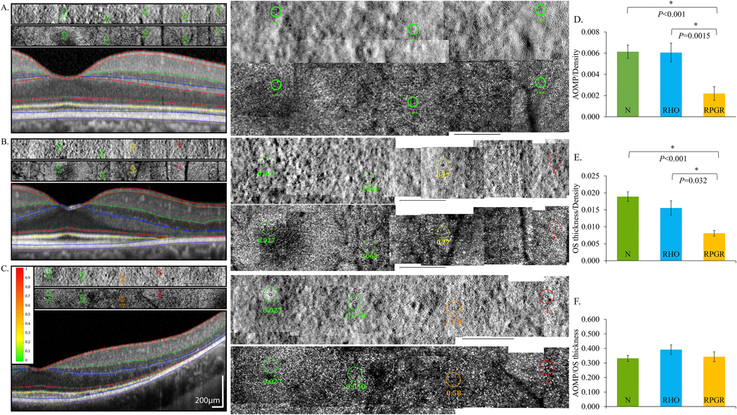
Adaptive optics microperimetry (AOMP) revealed reduced sensitivity per cone density in eyes with mutations in RPGR, which is expressed in rod and cone photoreceptors, compared to normal eyes and eyes with mutations in RHO, which is expressed exclusively in rods. AOSLO split detector (top) and confocal (center) images with test locations and sensitivities are shown as colored circles above a spectral domain OCT scan (bottom) from the same retinal location. Retinal sensitivities are displayed using a color scale ranging from green (normal) to red (not seen). Center panels show AOSLO images at 200% magnification; black scale bars in all AOSLO images are 1 degree. A: Sensitivities from a normal subject are normal, B: sensitivities from a patient with RHO c.810C>A, p.Ser270Arg are normal near the fovea but reduced (yellow) beginning at 4 degrees and not measurable (red) at 6 degrees temporal to the fovea, and C: sensitivities from a patient with RPGR c.1243_1244delAG, p. Arg415Glyfs*37 are normal near the fovea but more severely reduced (orange) beginning at 4 degrees and not measurable (red) at 6 degrees temporal to the fovea. D: Retinal sensitivity per cone density was significantly lower in eyes with RPGR mutations than normal eyes and eyes with RHO mutations. E: Outer segment (OS) thickness per cone density was significantly lower in eyes with RPGR mutations than eyes with RHO mutations and also significantly lower than normal. F: Retinal sensitivity was not significantly different among eyes with RHO and RPGR mutations or different from normal when normalized for OS thickness. Modified from Foote et al. (2020).
