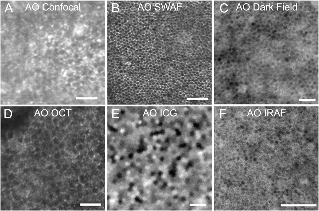
Fig. 5.
Images of retinal pigment epithelial (RPE) cells. A: RPE cells visualized in confocal adaptive optics scanning laser ophthalmoscopy (AOSLO) images of a patient with cone loss due to cone rod dystrophy (Roorda et al., 2007). B: AOSLO with short wavelength autofluorescence (AO-SWAF) capability shows RPE images in a monkey (Morgan et al., 2009a). C: AOSLO dark-field images show hexagonal RPE cells in a normal subject (Scoles et al., 2013). D: AO optical coherence tomography (AO-OCT) images of RPE cells (Liu et al., 2016). E: AO-indocyanine green (AO ICG) image of RPE cells (Tam et al., 2016). F: AO infrared autofluorescence (AO IRAF) images of RPE cells (Liu et al., 2017b). Scale bars, 50 μm. Modified from Roorda et al. (2007) and Morgan et al. (2009a), copyright by the Association for Research in Vision and Ophthalmology, Liu et al. (2016), Tam et al. (2016), Scoles et al. (2013) reprinted/adapted with permission from Drew Scoles, Yusufu N. Sulai, and Alfredo Dubra and Liu et al. (2017b), © The Optical Society.

