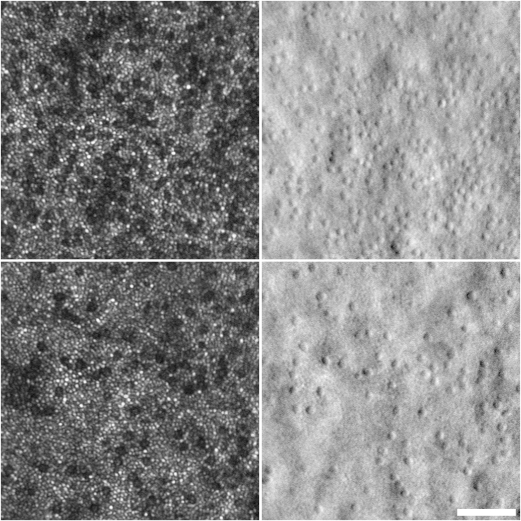
Fig. 8.

Confocal and corresponding split detection AOSLO images from one 43-year old male with CNGB3-associated ACHM. Dark areas on confocal imaging (left panels) correspond to cone inner segments on split-detection (right panels). The top and bottom image pairs are from 2.6 degrees and 4.6 degrees temporal to the fovea, respectively. Scale bar = 50 μm.
