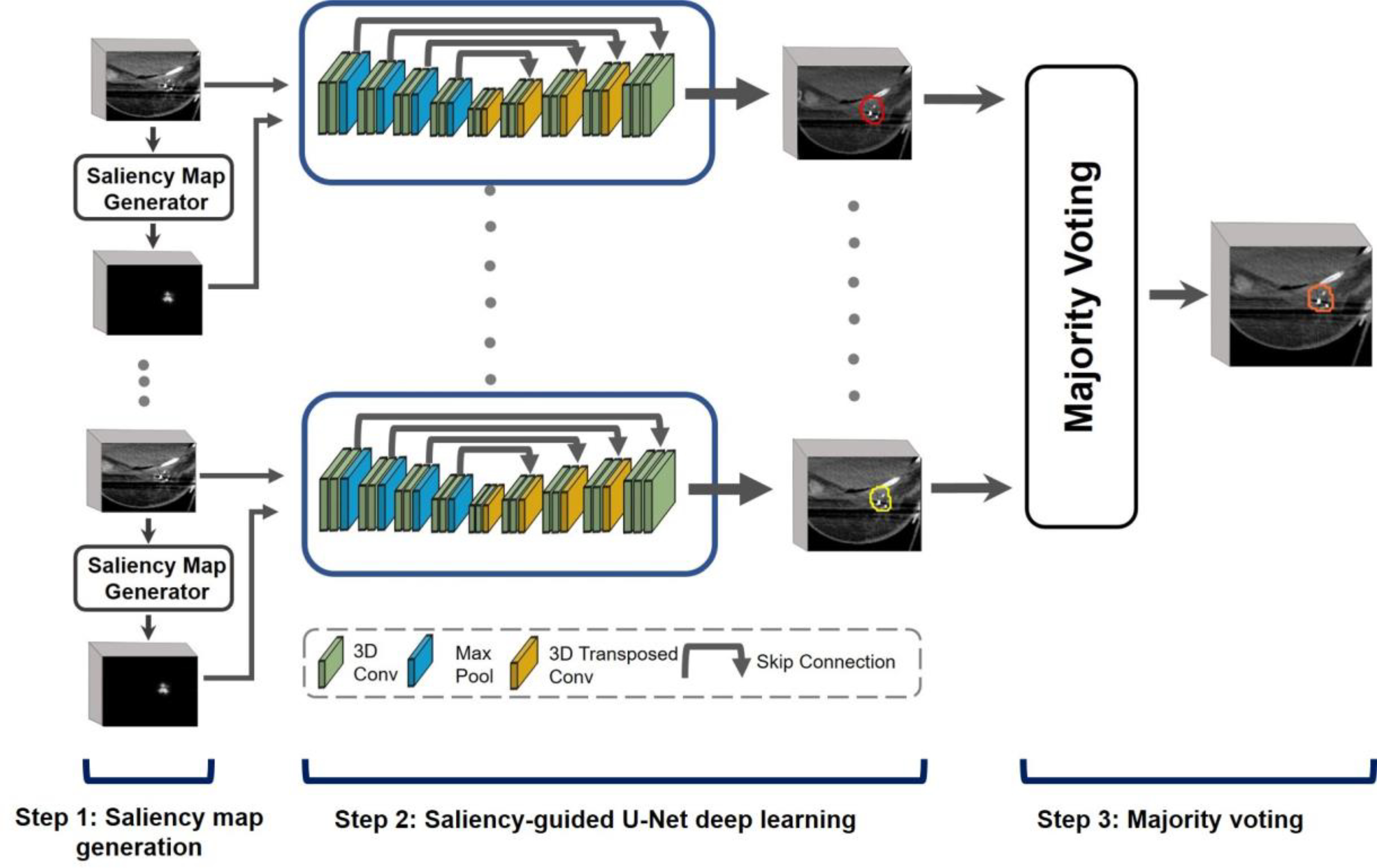
Figure 1.

Illustration of SDL-Seg structure. The model consists of three main steps: 1. Saliency map generation where saliency maps with target location cues are produced, 2. Saliency-guided U-Net encoding the regions with high saliency for more accurate segmentation of breast target. 3.Majority voting fuses predictions of multiple U-Nets to generate final segmentation
