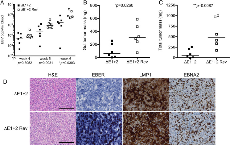
Fig. 2.
The M81 EBERs potentiate cell growth in vivo. (A) Primary B cells exposed to M81/ΔE 1+ 2 or M81/ΔE1 + 2 Rev were injected into NSG mice (n = 6 per group). Viral titers in the peripheral blood of infected mice were determined by qPCR at different weeks postinfection. One mouse from the M81/ΔE1 + 2 rev group developed a tumor and died between week 5 and week 6. Central horizontal lines represent the median. The results were analyzed with a Mann–Whitney U test. (B) The dot plot shows the gut tumor mass in animals that developed a tumor. Central horizontal lines represent median. The results were analyzed with a Mann–Whitney U test. (C) same as in B for the analysis of total tumor mass. (D) These pictures show immunohistochemistry stains of tumors that developed in the gut. (Scale bar, 100 µm.) Continuous tissue sections were stained with hematoxylin and eosin (H&E), immunostained with antibodies specific for LMP1, EBNA2, or subjected to an in situ hybridization with an EBER-specific probe.

