Fig. 1.
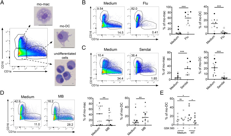
Pathogen recognition impacts monocyte differentiation. Monocytes were cultured for 5 d with M-CSF, IL-4, and TNF-α. Monocyte-derived cells were stained for CD16 and CD1a and analyzed by flow cytometry. (A) CD16+ mo-mac, CD1a+ mo-DC, and CD16-CD1a–undifferentiated cells were sorted and stained with May–Grunwald–Giemsa solutions after cytospin. (Scale bar, 30 μM.) Representative images (n = 5). (B–D) Proportions of mo-mac and mo-DC at day 5 after monocyte exposure to inactivated influenza A virus (Flu, B, n = 12), live Sendai virus (C, n = 8), and heat-killed mycobacterium butyricum (MB) (D, n = 15). Each symbol represents one individual donor. (E) Monocytes were preincubated with GSK583, heat-killed mycobacterium tuberculosis (MT) was added, and differentiation was analyzed after 5 d of culture. Wilcoxon test. *P < 0.05; **P < 0.01; ***P < 0.001; ns, not significant.

