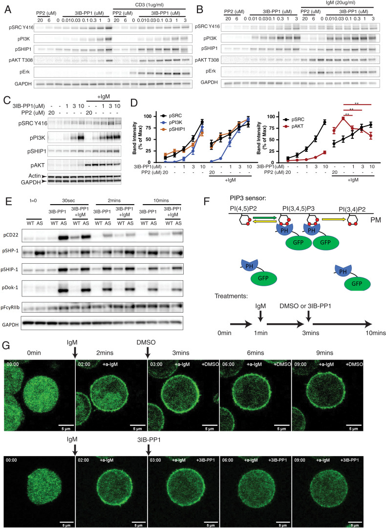
Fig. 3.
CskAS inhibition activates ITIM receptors and compromises AKT activation in B cells. (A–B) Double-positive thymocytes (A) and B cells (B) isolated from CskAS mice were stimulated with indicated reagents for 2 min. (C and D) BAL17-CskAS cells were stimulated with 10 μg/mL F(ab′)2 anti-μ (IgM) and indicated reagents for 2 min. (C) Phosphorylation of indicated molecular targets was assessed by immunoblots. (D) The band intensities in C were quantified and normalized to the band with highest intensity of each molecular target. **P < 0.01 (one-way ANOVA test with Dunnett’s multiple comparisons test, n = 8, error bars, SEM). (E) B cells from wild-type (WT) and CskAS (AS) mice were stimulated similarly as in Fig. 1A. Phosphorylation of selected ITIM receptors and associated phosphatases were assessed by immunoblots. (F and G) BAL17-CskAS cells were transfected with a PIP3 GFP biosensor. The cells were stimulated with 20 μg/mL F(ab′)2 anti-μ (IgM) and 10 μM 3IB-PP1 or DMSO for the indicated time intervals (F). The translocation of the PIP3 sensor was imaged by confocal microscopy continuously for 10 min. Selected key frames are shown (G). Full movie images are presented in Movies S1 and S2. All data are representative of at least two independent experiments.

