Fig. 2.
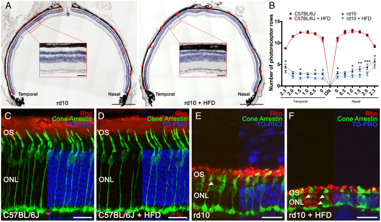
High-fat intake accelerates photoreceptor cell degeneration. (A) Representative nasal–temporal retina cross-sections and high magnification views from rd10 mice fed a normal or HFD for 2 wk from postnatal day 19 stained with hematoxylin. Red marks point to regions where the number of photoreceptor rows was quantified. (B) Average number of photoreceptor rows throughout the nasal–temporal axis in C57BL/6J and rd10 mice fed normal or HFD (n = 3 for C57BL/6J mice and n = 8 to 9 for rd10 mice). (C–F) Representative retina cross-sections from C57BL/6J and rd10 mice fed normal chow or HFD immunolabeled against rhodopsin (Rho, rod cells, in red) and cone arrestin (cone cells, in green). TO-PRO 3– (in blue) stained nuclei. In C57BL/6J mice, images show a normal cone photoreceptor morphology and rhodopsin distribution (C and D), independently of the diet. In rd10 mice, images show morphological alteration of the cones and abnormal distribution of rhodopsin (E), both aggravated by HFD consumption (F). Arrowheads point to the mislocalization of rhodopsin into the cell bodies (E and F). ANOVA, Bonferroni’s test, diet effects (normal versus HFD), *P < 0.05, **P < 0.01, ***P < 0.001. OS: outer segment. (Scale bars: A, 500 μm; Insets, 50 μm; C–F, 20 μm.)

