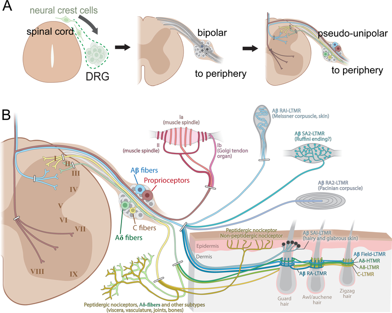
Figure 1. Overview of early development, anatomy, and the major DRG somatosensory neuron subtypes.
(A) Dorsal root ganglia (DRG) neurons derive from neural crest cells that delaminate from the dorsal neural tube and coalesce to form DRGs. Rodents typically have 30 or 31 pairs of DRGs; 8 pairs of cervical, 13 pairs of thoracic, 5 or 6 pairs of lumbar, and 4 pairs of sacral DRGs. Nascent DRG neurons assume a spindle-shaped, bipolar morphology, with axons emanating from opposite sides of the cell body. Then, a stem axonal protrusion containing the two axonal branches forms and refines to assume the mature T shape pseudo-unipolar morphology by birth.
(B) Somatosensory neuron cell bodies reside in DRG, adjacent to the spinal cord. DRG neurons have a pseudo-unipolar morphology with axonal branches extending into both the periphery and the spinal cord. The major peripheral end organs formed by DRG neuron subtypes are illustrated on the right, and the general, albeit oversimplified spinal cord lamination patterns of their central projections are also shown. Note that Aβ LTMR fibers and proprioceptors also have central branches with multiple collaterals extending along the rostral-caudal axis of the spinal cord and an additional branch that ascends via the dorsal column, often reaching the dorsal column nuclei of the brainstem.

