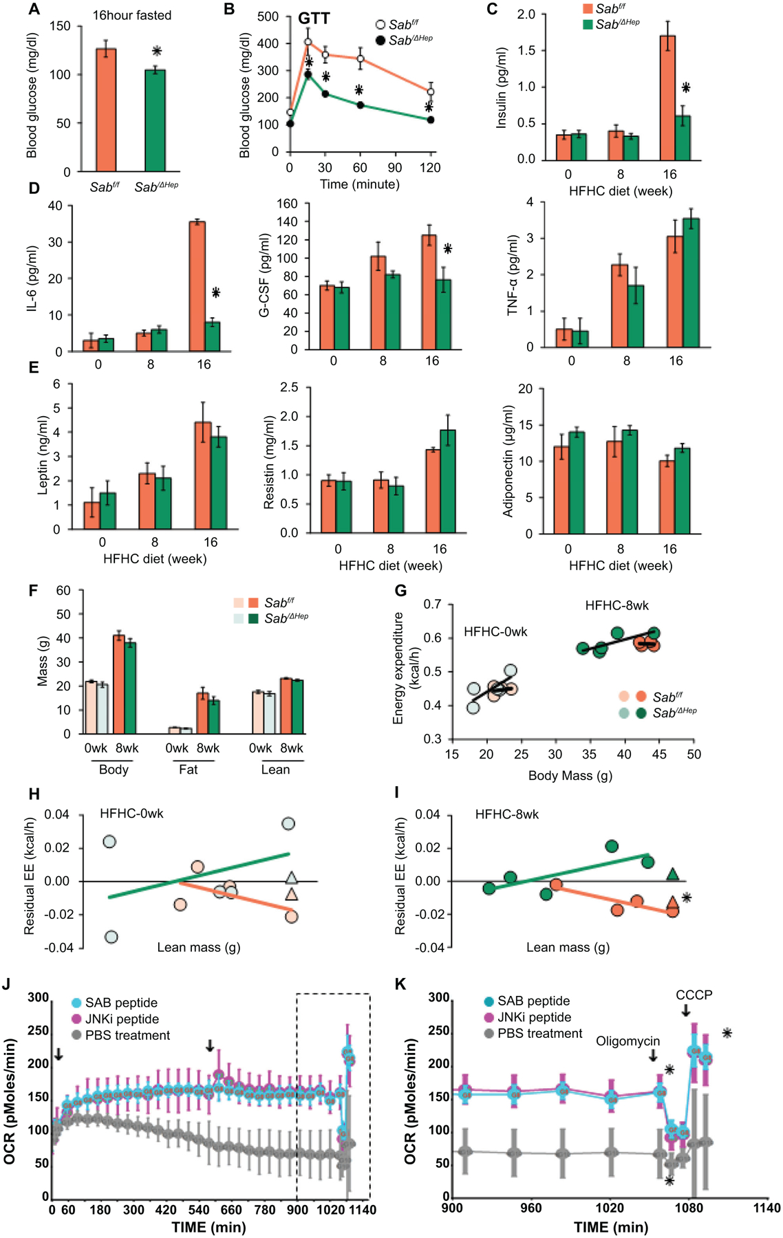
Fig.3. Sab deletion prevents diet-induced metabolic intolerance and inflammatory response.

Male Sabf/f and SabiΔHep mice were fed the HFHC diet up to 16 weeks. (A) 16 hours fasting blood glucose, (B) glucose tolerance test (GTT) (C) serum insulin after 4 hours fast. (D) Serum inflammatory cytokines: IL-6, G-CSF, TNF, and (E) serum leptin, resistin, and adiponectin in 4 hours fasted mice. All data are presented as mean ± SEM. n=5 mice/group, ✱=p<0.05 SabiΔHep versus Sabf/f by unpaired, 2-tailed Student’s t-test. (F) Body, fat and lean mass, n=5 mice per group. (G) Energy expenditure (EE) measured by PhenoMaster/LabMaster home cage System. (H,I) Residual EE obtained from regression plot was graphed against lean mass. Mean value of residuals of EE are shown as (Δ). Statistical significant difference (✱=p<0.05) of EE in SabiΔHep versus Sabf/f treatment by ANCOVA. n=4–5 mice/group. (J) Lipid metabolism of steatotic hepatocytes examined by Seahorse XF analyzer. PMH from wild type C57BL/6N mice fed HFHC diet for 12 weeks were seeded on collagen coated Seahorse cell culture plate. Cells were washed in serum, glucose and pyruvate-free-XF medium. After measurement of basal level, SAB-peptide or JNKi-peptide (5 μM) was injected. OCR was measured over the time course (18 hours) with second injection (arrow) 9 hours after first injection. (K) Final 3 hours measurements (boxed in J) showing the oligomycin inhibitable (ATP coupled) respiration and CCCP induced maximal (uncoupled) respiration. n=3 separate experiments, ✱=P<0.05 peptide versus vehicle treatment by two-way ANOVA. Data represents mean ± SEM.
