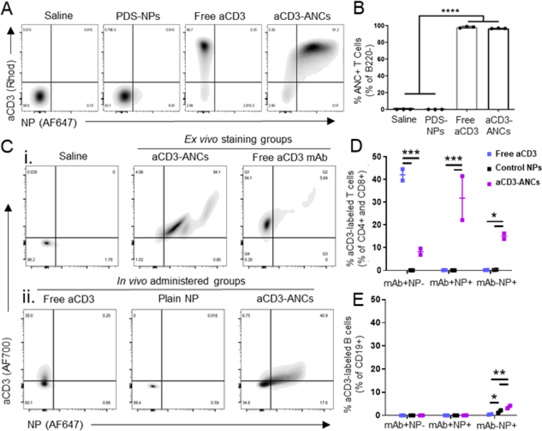
Figure 2: ANCs retain mAb binding in vitro and in vivo.

A) Representative flow cytometry plots of binding by various aCD3 mAb (AlexaFluor700-labeled) and NP (AlexaFluor647-labeled) formulations to B220− murine splenocytes after ex vivo co-incubation for 15 min. B) Quantification of NP+ (AlexaFluor647+) B220− murine splenocytes by various aCD3 mAb (AlexaFluor700-labeled) and NP (AlexaFluor647-labeled) formulations after ex vivo co-incubation for 15 min, as shown in A. C) Flow cytometry plots of aCD3 mAb (AlexaFluor700-labeled) and NP (AlexaFluor647-labeled) association with LN-resident CD4+ & CD8+ T cells after ex vivo incubation with free or ANC-formulated aCD3 (i) or in vivo 24 h post i.d. injection of free aCD3, unconjugated PDS-NPs, or aCD3-ANCs in naïve mice (6.25 μg total mAb dose) (ii). Quantified in vivo flow cytometry data from Cii. Frequencies represent the proportion of total CD4+ and CD8+ (D) or CD19+ (E) cells in each subgate: mAb+NP− (AF700+AF647−), mAb+NP+ (AF700+AF647+), and mAb−NP+ (AF700−AF647+). Statistical analyses were done using one- (B) or two-way (D-E) ANOVA with Tukey’s test. *p<0.05, **p<0.01, ***p<0.001, ****p< 0.0001.
