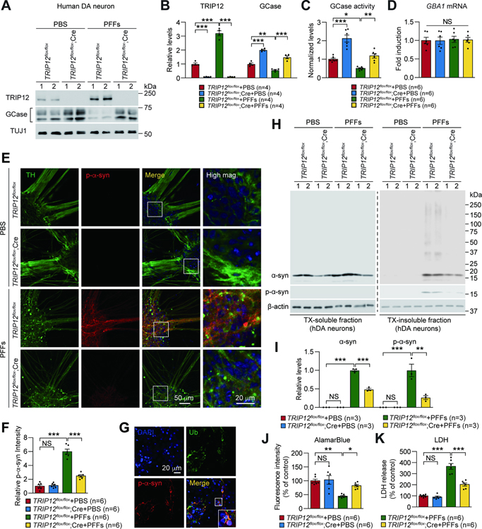
Figure 5. TRIP12 Knockout Rescues α-syn PFFs-induced Pathologies in Human Dopaminergic (DA) Neurons.
(A) Western blot analysis of TRIP12 and GCase in conditionally floxed TRIP12 hESCs-derived human dopaminergic (DA) neurons transduced with AAV expressing either control (TRIP12flox/flox) or cre recombinase (TRIP12flox/flox;Cre) and treated with PBS or α-syn PFFs.
(B) Data in A shown as bar graphs (n=4, each group).
(C) GCase activity (n=6, each group).
(D) GBA1 mRNA level (n=6, each group).
(E) Representative confocal images showing p-α-syn aggregates.
(F) Data in E shown as bar graphs (n=6, each group).
(G) The p-α-syn positive signals co-localize with ubiquitin.
(H) Western blot analysis of α-syn, p-α-syn, and β-actin from TX-soluble and TX-insoluble fractions.
(I) Bar graph of the levels of α-syn aggregates and p-α-syn in TX-insoluble fraction (n=3, each group).
(J) Quantification of AlamarBlue assay (n=6, each group).
(K) LDH assay (n=6, each group). Data are presented as mean ± SEM (NS; not significant, *P < 0.05, **P < 0.01, ***P < 0.001).
See also Figure S6.

