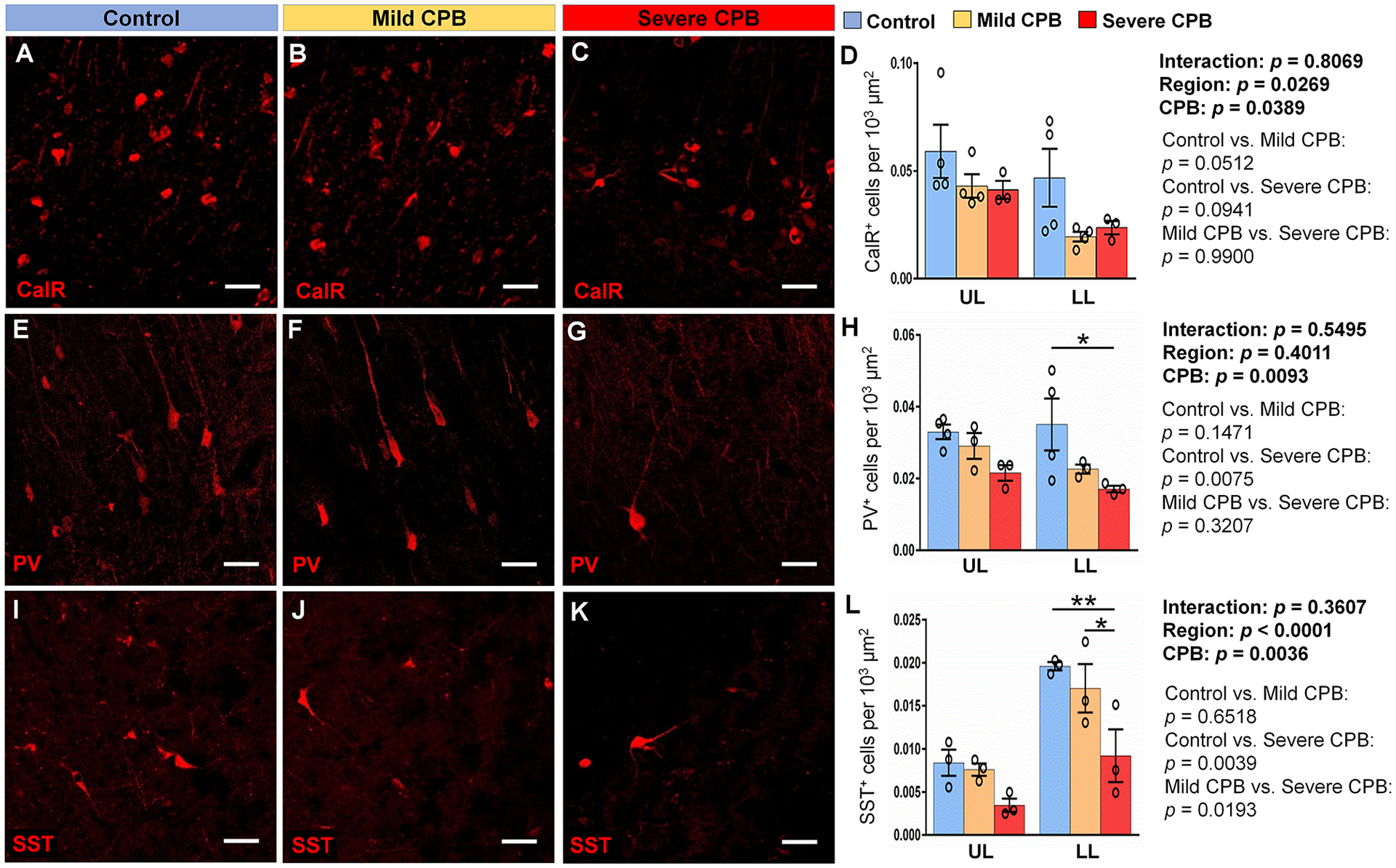
Figure 5. CPB alters interneuron cell populations in the cortex 4 weeks post-surgery.

(A-C) Immunostains of Calretinin within the lower cortical layers of the frontal lobe. (D) Quantification of the average density of CalR+ cells. (E-G) Immunostains of Parvalbumin within the lower cortical layers of the frontal lobe. (H) Quantification of the average density of PV+ cells. (I-K) Immunostains of Somatostatin within the lower cortical layers of the frontal lobe. (L) Quantification of the average density of SST+ cells. Scale bars, 50 μm. Data are expressed as mean ± SEM (n = 3 to 4 animals per group). *p <0.05, **p < 0.01, two-way ANOVA with Tukey’s post-hoc test (D, H, L). CalR, calretinin; CPB, cardiopulmonary bypass; FL, frontal lobe; LL, lower layers; PV, parvalbumin; SST, somatostatin; UL, upper layers.
