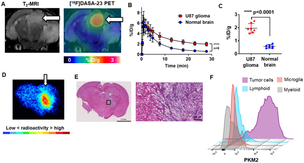
Figure 2.
Evaluation of [18F]DASA-23 in orthotopic U87-GFP/luc GBM mice. A Representative images of a mouse bearing an orthotopic U87-GFP/luc GBM (white arrow), the presence of the tumor is confirmed with T2-weighted MRI. A representative [18F]DASA-23 fused PET/CT/MR image is shown summed 10-30 min post tracer administration. B Time activity curves illustrating the uptake and clearance of [18F]DASA-23 over time in the U87-GFP/luc GBM and contralateral normal brain, p<0.0001, n=8. C Quantification of [18F]DASA-23 radioactivity in U87-GFP/luc GBM and contralateral normal brain at 30-min post tracer administration, t-test, p<0.0001, n=8. D Representative [18F]DASA-23 autoradiography in mice bearing orthotopic U87-GFP/luc GBM at 30-mins post tracer administration. E H&E stain of an adjacent section showing the presence of the highly cellular GBM. F Expression profile of PKM2 in the brain of mice bearing orthotopic U87-GFP/luc GBM using flow cytometry (n=4).

