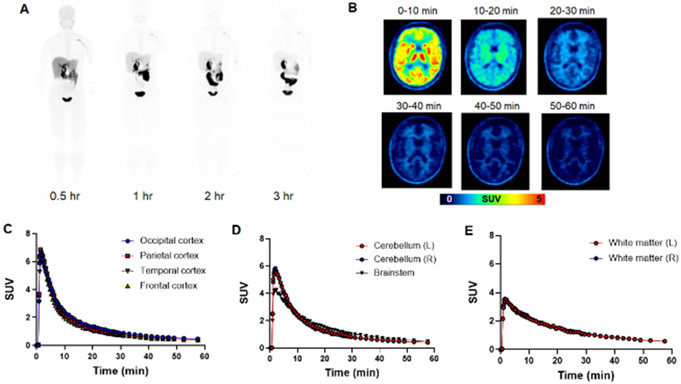
Figure 3.
Evaluation of [18F]DASA-23 in healthy human volunteers. A Representative [18F]DASA-23 whole body maximum intensity projections show the biodistribution of the PET tracer at various time points post tracer administration in volunteer 3. B Representative axial [18F]DASA-23 PET images of a healthy human brain at various summed time points post tracer administration. C Time activity curves showing [18F]DASA-23 uptake and clearance in the cerebral cortices, D posterior fossa and E white matter in the left (L) and right (R) brain hemispheres.

