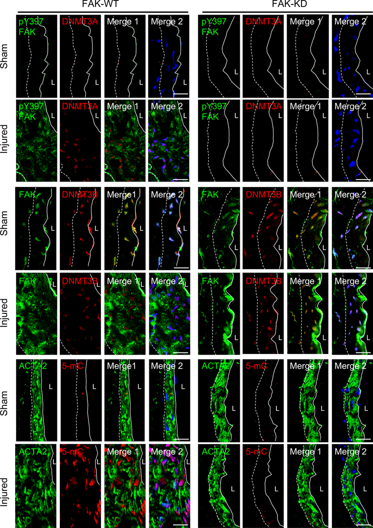
Figure 6. SMC-specific genetic FAK-KD mice did not upregulate injury-induced DNMT3A and 5-mC expression protecting from neointima formation.
Representative immunofluorescence staining of FAK-WT and FAK-KD femoral arteries 2 weeks postinjury for FAK, pY397 FAK, DNMT3A, DNMT3B, 5-mC and ACTA2 (n=4). FAK-KD in SMCs from the injured group exhibited a low DNMT3A and 5-mC as observed in sham. Merge 1, green and red; Merge 2, green, red, and blue (DAPI) were merged. Dashed line in image marks the elastic lamina. White line indicates endothelial layer. L, lumen. Scale bars: 20 μm.

