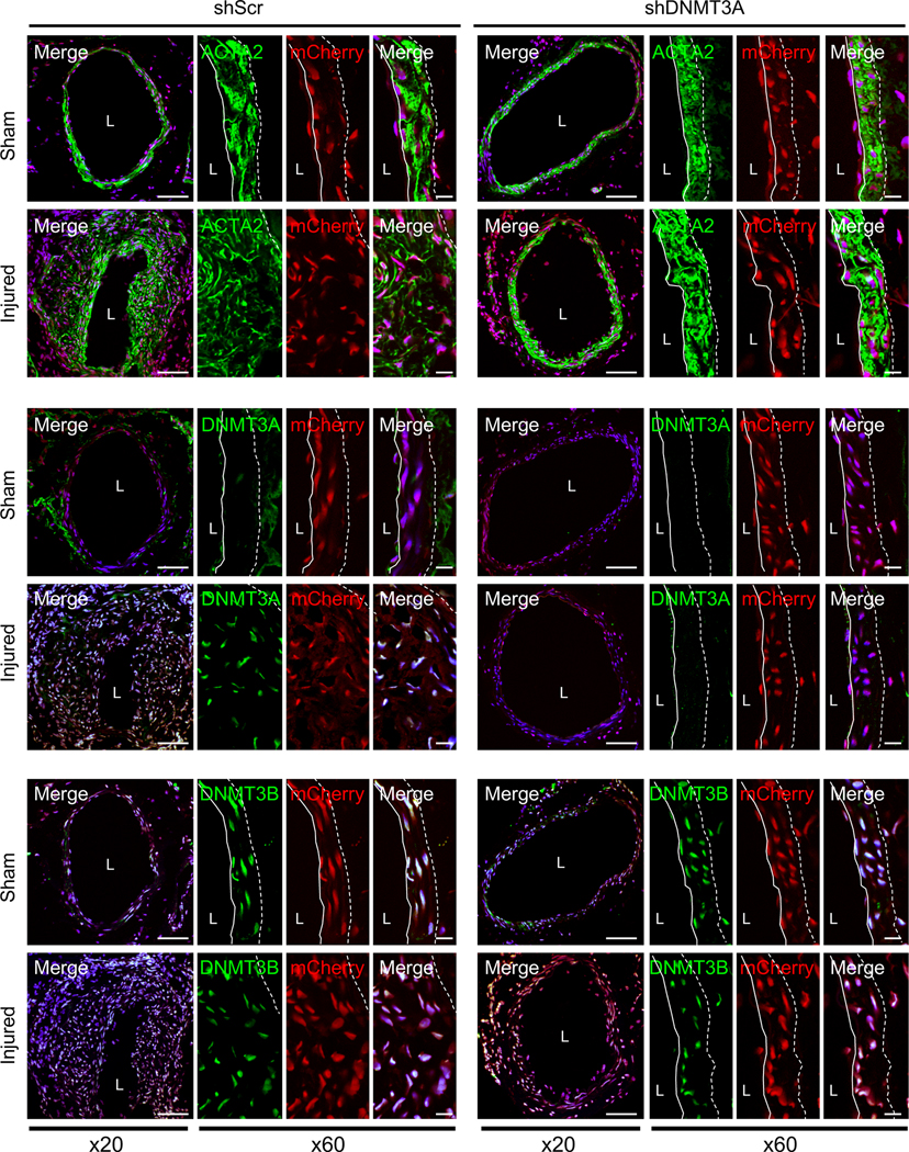
Figure 8. In vivo knock-down of DNMT3A in femoral artery reduces wire injury-induced neointima formation.
Femoral arteries were coated with either scramble shRNA (shScr) or DNMT3A shRNA (shDNMT3A) lentivirus coexpressing mCherry immediately following wire injury. Representative immunostainings of 2-week post-injury samples for ACTA2, DNMT3A and DNMT3B are shown (n=4). Green, mCherry (red) and DAPI (blue) were merged. mCherry was used to verify lentiviral infection. Dotted line in image marks the elastic lamina. White line indicates endothelial layer. L, lumen. Scale bars: x20, 100 μm; or x60, 20 μm.

