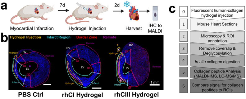
Figure 1.
Mouse model study design and multimodal imaging mass spectrometry workflow. (a) Timeline of mouse model. 9-week old mice were induced with myocardial infarction. 7-days postsurgery, collagen hydrogel or PBS control was injected into the left ventricle. 2-days post injection, hearts were harvested. (b) Fluorescence microscopy images showing region of interest (ROI) annotations of heart sections in relation to fluorescently labeled human hydrogel signal. BZ: border zone; LV: left ventricle; RV: right ventricle. Scale bar shown is 1 mm. (c) Simplified workflow of tandem high-resolution fluorescent microscopy and collagenase-based MALDI IMS and LC MS/MS experiments.

