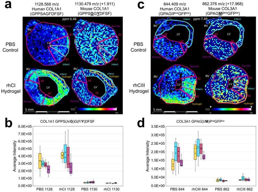
Figure 3.
Collagen peptides unique to human proteome have differential distribution to corresponding mouse peptides. Species differentiating amino acids are underlined in bold within the sequence. (a) Representative collagen type 1 peaks as visualized in rhCI injected hearts as compared with PBS controls are shown for the human-specific collagen sequence (left) and corresponding mouse sequence (right). Average intensity levels measured between all samples studied are shown in (b). (c) Representative collagen type 3a1 peaks as visualized in rhCIII injected hearts as compared to PBS controls are shown for the human-specific collagen sequence (left) and corresponding mouse sequence (right). Average intensity levels measured between all samples studied are shown in (d). n = 3. Six tissue sections were visualized for each biological replicate. Different representative samples are used to show reproducibility across n’s. Note: Location of hydroxyproline (Pox) are ambiguous and require further validation. Peptides shown in a–d represent nearest identified putative peptides from previously acquired human databases.

