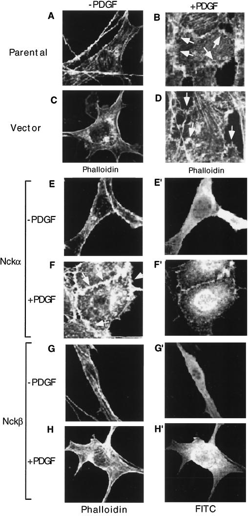
FIG. 1.
Overexpression of Nckβ but not Nckα blocks PDGF-stimulated membrane ruffling. NIH 3T3 cells, cultured in fibronectin-coated (10 μg/ml, 2 h) eight-chamber culture slides, were either untransfected (A and B), transfected with vector alone mixed with a GFP-containing vector (C and D), or transfected with the wild-type Nckα (E to F′) or Nckβ (G to H′) construct at 0.5 μg/well. After 48 h, cells were starved in low-serum medium for an additional 18 h and treated (B, D, F, and H) or not (A, C, E, and G) with PDGF-bb (100 ng/ml) at 37°C for 15 min. Expression of the Nck proteins was monitored by anti-HA antibody blotting, followed by a secondary antibody conjugated with FITC. The actin cytoskeleton was revealed by rhodamine-labeled phalloidin staining. Eighty to 100 cells which showed positive FITC staining were selected and analyzed in each experiment. Vector-transfected cells were identified as GFP positive. Images were recorded with a Zeiss confocal microscope. Magnifications, × 150. This experiment was repeated four times.

