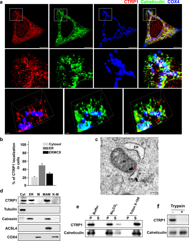
Fig. 1. CTRP1 is an ER membrane protein.
a Representative low-magnification images (top), high-magnification images of the boxed areas (middle) and 3D reconstruction images (bottom) of mouse embryonic fibroblasts (MEFs) immunostained with CTRP1 (red), the ER marker calreticulin (green), and the mitochondrial protein COX4 (blue) antibodies. The white arrows indicate CTRP1 in the EMCSs. Scale bars, 10 µm (top) and 1 µm (bottom). b Quantitation of CTRP1 localization to the cytosol, ER and ERMCSs in MEFs. c Representative electron micrograph of MEFs stained with anti-CTRP1 conjugated to 10-nm gold nanoparticles (red arrows). M mitochondrion, ER endoplasmic reticulum. Scale bar, 200 nm. d Western blot analysis of subcellular fractions of MEFs. Cyt cytosol, MAM mitochondria-associated ER membrane, K-M Kit-purified mitochondria. The subcellular fractions were immunoblotted with antibodies against the cytosolic marker tubulin, the ER marker calnexin, the MAM marker ACSL4, and the mitochondrial marker COX4. e Western blot analysis of the pellet (P) and supernatant (S) fractions collected after ER samples were incubated with buffer, Na2CO3 or Triton X-100. f Western blot analysis of ER fractions that were treated with or without trypsin. Throughout, the data are presented as the mean ± s.e.m.

