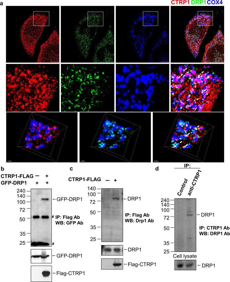
Fig. 3. CTRP1 interacts with DRP1.
a Representative low-magnification images (top), high-magnification images of the boxed areas (middle) and 3D reconstruction images (bottom) of MEFs immunostained with antibodies against CTRP1 (red) and DRP1 (green) and the mitochondrial protein COX4 (blue). The white arrows indicate colocalization of CTRP1 and DRP1. Scale bars, 1 µm (top) and 10 µm (bottom). b Whole-cell lysates (WCLs) of 293T cells cotransfected with constructs expressing Flag–CTRP1 and GFP–DRP1 were immunoprecipitated (IP) with anti-Flag beads and then immunoblotted (IB) with a GFP-specific antibody. Asterisks indicate immunoglobulin G (IgG). c Interaction between endogenous DRP1 and Flag-CTRP1; 293T cells were transfected with Flag–vector (Lane 1) or Flag–CTRP1 (Lane 2), and WCLs were immunoprecipitated with anti-Flag beads and blotted with anti-DRP1. d Interaction between endogenous DRP1 and CTRP1. WCLs of 293T cells were immunoprecipitated with anti-CTRP1 and blotted with anti-Drp1.

