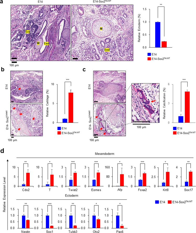
Fig. 5. Teratomas derived from the E14-Sox2TA/WT cells exhibited decreased ectodermal lineage commitment and increased cartilage formation.
a The E14 and E14-Sox2TA/WT cells were transplanted into nude mice. Teratomas that formed 14 days after transplantation were analyzed by hematoxylin and eosin staining. The E14-Sox2TA/WT cells exhibited decreased ectodermal lineage commitment. Scale bars, 100 μm. Ect ectodermal lineage, M mesodermal lineage, End endodermal lineage. The degree of ectodermal lineage commitment was quantified using InForm 2.4.10 image analysis software (PerkinElmer), and the relative amounts of ectoderm formed (%) are presented as the mean ± standard deviation (n = 3). b The E14-Sox2TA/WT cells exhibited increased cartilage formation. Representative images and the quantification results are shown. Red arrows indicate cartilage. c The E14-Sox2TA/WT cells exhibited increased calcification. Representative images and the quantification results are shown. Red arrows indicate calcified tissue. d The mRNA expression levels of mesendodermal and ectodermal markers in teratomas were analyzed using real-time qPCR. The graph shows the relative expression levels (mean ± standard deviation, n = 3) after normalization to ActB expression. *P < 0.05, **P < 0.01, ***P < 0.001.

