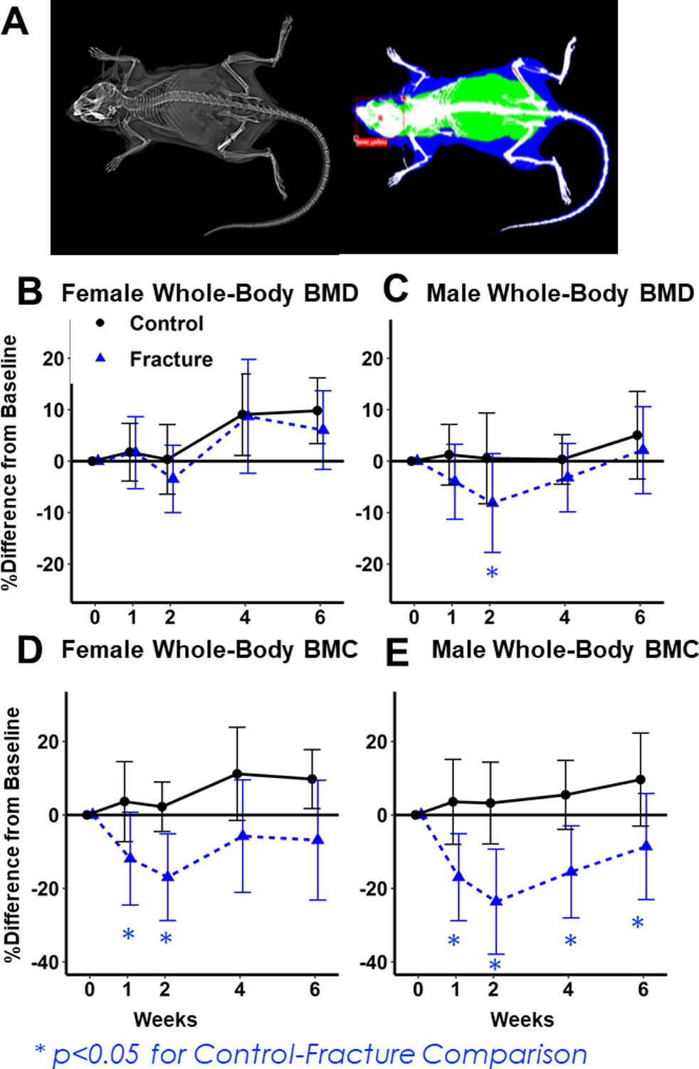
Figure 1: Dual-energy X-ray absorptiometry (DXA) imaging of whole-body BMD and BMC.

(A) Representative image for DXA analysis. (B, C) Percent change from baseline in Females (B) and Males (C) BMD. Decreases in Fracture mice relative to Controls were greater in Males. (D,E) Percent change from baseline in Female (D) and Male (E) BMC. Percent decreases were greatest in Male Fracture mice.
