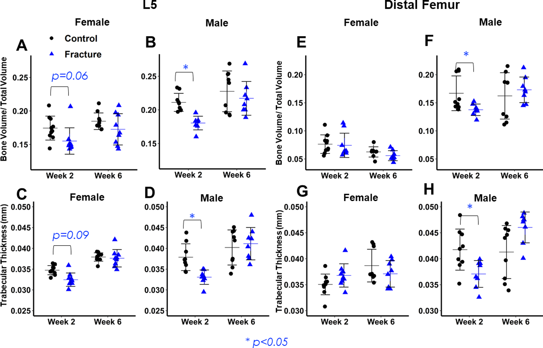
Figure 3: Micro-computed tomography analysis of 5th lumbar vertebrae (L5) and distal femur trabecular bone.

(A,B,C,D) L5 microarchitectural properties. Lower values of bone volume/ total volume (A,B) and trabecular thickness (C,D) in Fracture mice are evident in both sexes at 2 weeks post-fracture, but greater in Males. No differences between Fracture and Control mice 6 weeks post-fracture. (E,F,G,H) distal femur microarchitectural properties. Lower values of bone volume/ total volume (E,F) and trabecular thickness (G,H) in Fracture mice are evident in Males 2 weeks post-fracture, and there are no differences between Fracture and Control mice at 6 weeks post-fracture.
