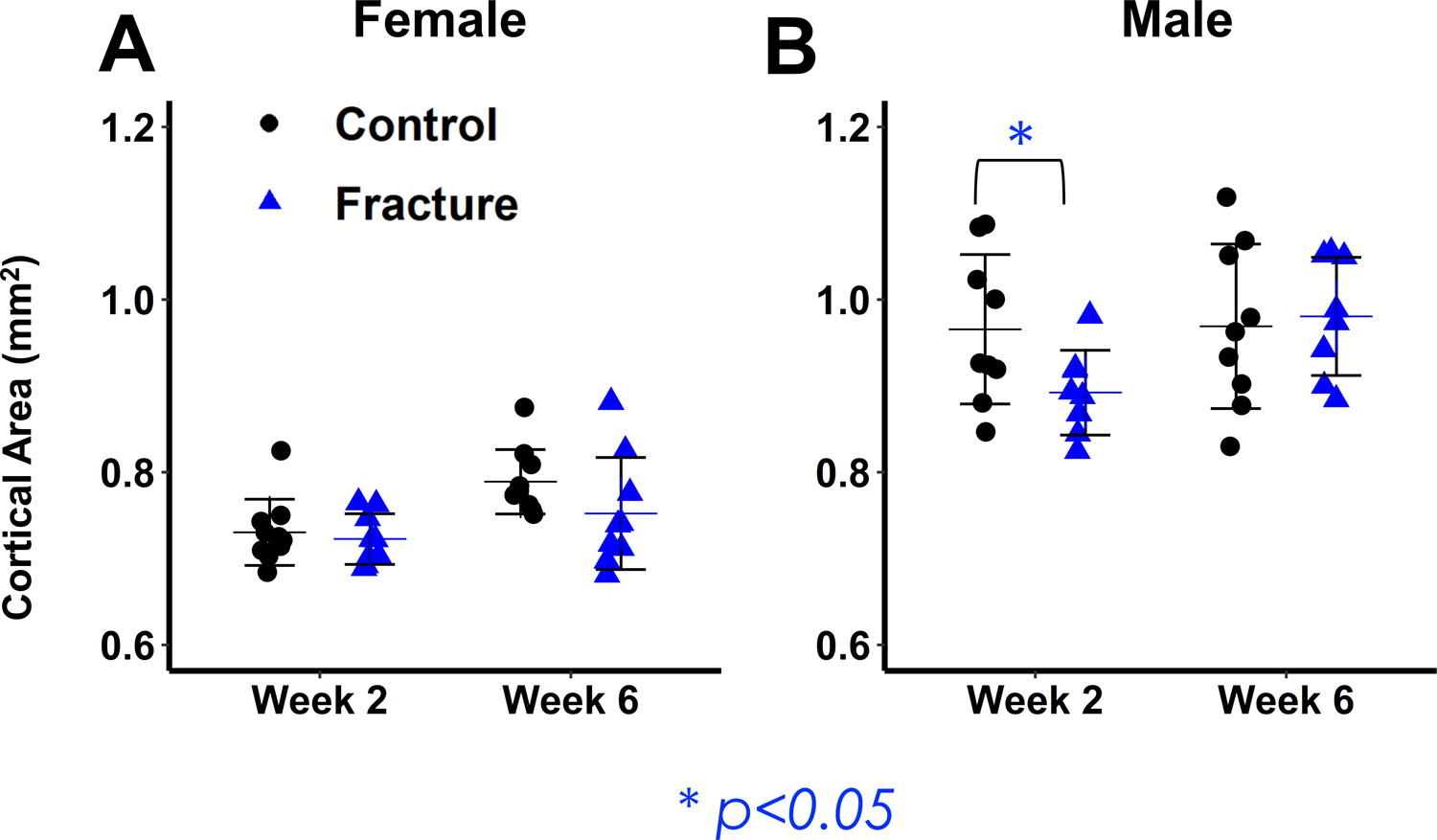
Figure 4: Micro-computed tomography analysis of femur midshaft cortical bone.

(A,B) Cortical Area. Male Fracture mice show significant decreases in cortical area at 2 weeks post-fracture and recover by 6 weeks post-fracture.

(A,B) Cortical Area. Male Fracture mice show significant decreases in cortical area at 2 weeks post-fracture and recover by 6 weeks post-fracture.