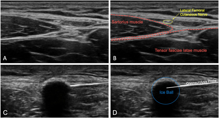
Figure 1.
(A) The lateral femoral cutaneous nerve is identified using ultrasound in the intermuscular plane between the sartorius and tensor fasciae latae muscles. (B) The lateral femoral cutaneous nerve (yellow) and sartorius and tensor fasciae latae muscles (red) are labeled. (C) Ultrasound is used to visualize the ice ball completely enveloping the lateral femoral cutaneous nerve, which can no longer be distinguished in the frozen tissue. (D) The sartorius muscle, cryoneurolysis probe, and ice ball are labeled.

