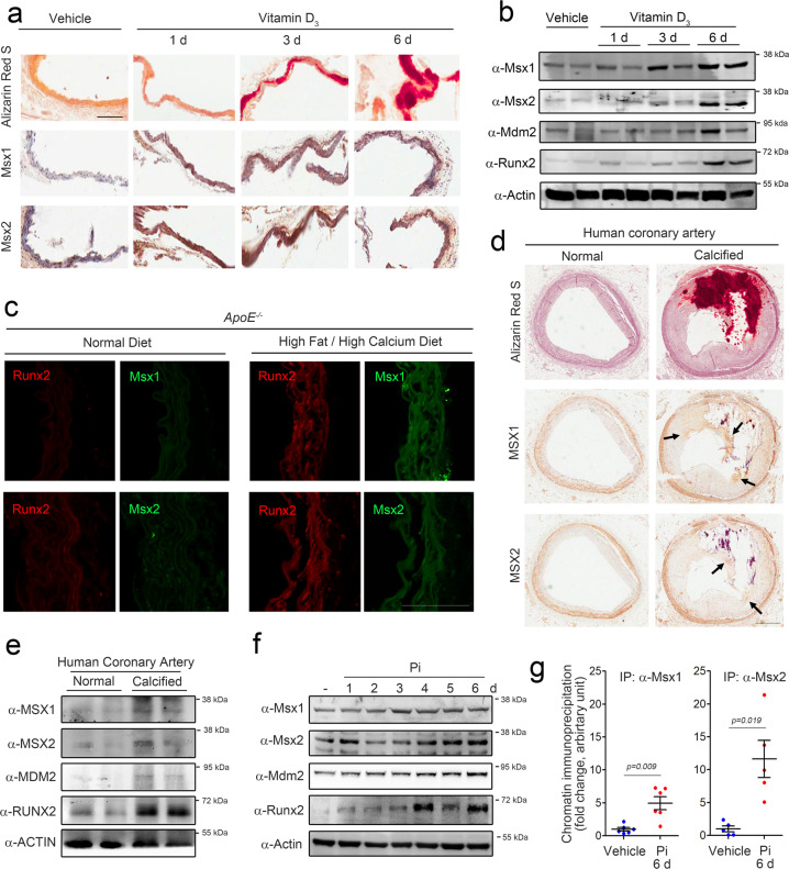
Fig. 4. MSX1 and MSX2 expression is induced in calcified vessels.
a Expression of Msx1 and Msx2 in aortas obtained from vitamin D3-treated mice (n = 4). Scale bar = 100 μm. b Vitamin D3 administration increased the protein levels of Msx1 and Msx2. c Expression of Msx1 and Msx2 in an alternative vascular calcification model induced in ApoE−/− mice (n = 3) by feeding a high-fat/high-calcium diet for 16 weeks. Scale bar = 100 μm. d Immunohistochemical analysis of human calcified coronary artery samples (n = 3). Scale bar = 500 μm. e Western blot analysis of human coronary artery samples. f Pi increased the protein levels of Msx1 and Msx2 in rat VSMCs. g Chromatin immunoprecipitation analysis showing the binding of Msx1 and Msx2 to MSXE in the MDM2 promoter in rat VSMCs in the presence of Pi.

