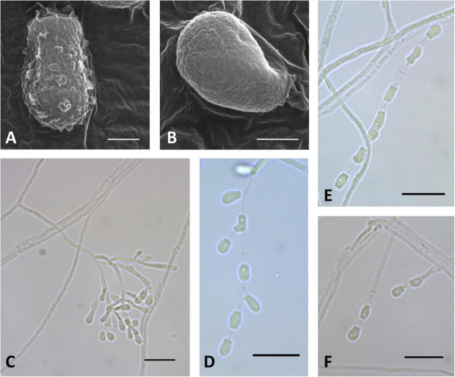
FIGURE 5.
Microscopic analysis of Pseudogymnoascus australis sp. nov. Conidia (A,B); Conidiophore (C); Fertile hyphae bearing arthroconidia and aleurioconidia, sessile, or stalked (D,E,F). In panels (A,B), the structures were observed using transmission electron microscopy, while in panels (C–F), light microscopy was used. Scale bars = 1 μm (A,B), 10 μm (C–F).

