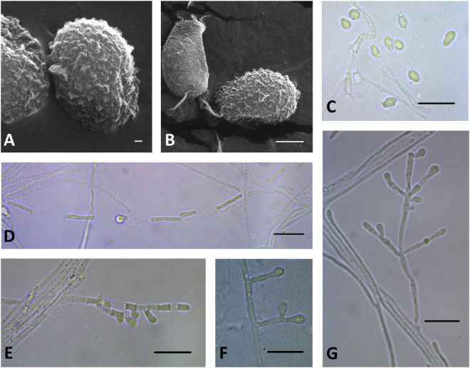
FIGURE 9.
Microscopic analysis of Pseudogymnoascus lanuginosus sp. nov. Conidia (A,B,C); Chain of arthroconidia (D); Fertile hyphae bearing arthroconidia and aleurioconidia, sessile, or stalked (E); Stalked aleurioconidia (F); Conidiophore (G). In panels (A,B), the structures were observed using transmission electron microscopy, while in panels (C–G), light microscopy was used. Scale bars = 200 nm (A), 1 μm (B), and 10 μm (C–G).

