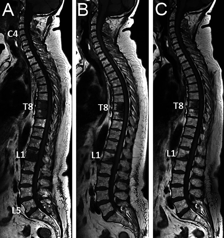
Figure 1.
53 year-old woman with newly diagnosed metastatic breast cancer (grade II ductal carcinoma, ER 8, PR8, KI 67 5%, HER2 neu 2+): spinal MRI findings at diagnosis of bone metastases and during treatment. (A) Baseline sagittal T1-weighted MR image of the whole spine shows multiple foci of low signal intensity of the bone marrow, typical for bone metastases (posterior arch of C4, vertebral bodies of T8, T9, L1, tiny foci in L5). (B) Corresponding MR image obtained 2-m later after combined treatment including a selective estrogen receptor degrader (SERD) and palbociclib shows significant decrease in size of all lesions, and disappearance of the small L5 foci. (C) Follow-up MR image obtained 2-m later shows further decrease in size of all lesions, with measurable decrease in lesion size and reappearance of fatty marrow at the periphery and within the lesions, again indicating frank response to treatment.

