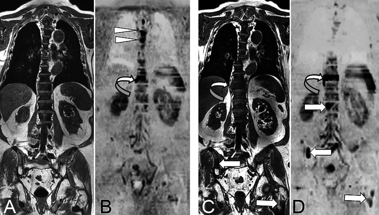
Figure 2.
73 year-old man with advanced prostate cancer. Comparison of pre- and post-treatment (enzalutamide) WB-MRI/DWI. Baseline coronal T1-weighted MR image (A) shows diffuse bone marrow infiltration within the spine, responsible for diffuse low signal intensity of the bone marrow, and related to advanced metastatic disease after several lines of treatment. The pelvic bones show higher signal of the bone marrow indicating a fatty content due to previous irradiation. Several focal lesions of low signal intensity are visible within the pelvis and left proximal femur. Baseline DWI MR image (B; B = 1000 s/mm2, inverted grey scale) shows high signal intensity foci typical for active bone metastases within the T4, T5 (arrowheads) and T10 (curved arrow) vertebrae. Follow-up T1-weighted MR image (C) shows no evident change of the spinal bone marrow, but increase in the right paraspinal extension of the T10 metastasis (curved arrow), and a new lesion within the right posterior iliac crest (arrow). Follow-up DWI MR image (D) shows disappearance of the midthoracic vertebral lesions, but increase in size and right paraspinal extension of the T10 vertebral lesion (curved arrow), and appearance of new lesions within the L1 vertebral body, the right iliac crest and left proximal femur (arrows). The observation of concurrent signs of disease response and progression is frequent, especially in advanced stages of metastatic cancer.

