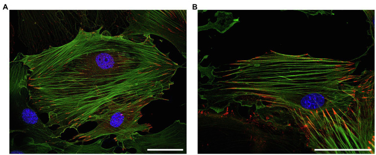
Figure 3.
Representative immunofluorescence analysis of the intracellular localization of (top) LPP and (bottom) zyxin in cultured vascular SMC isolated from the aorta of 3-month old C57BL/6 wildtype mice. The vascular SMC (passage 3) were seeded directly on microscopic slides, fixed with p-formaldehyde, and stained for LPP or zyxin with a corresponding anti-LPP (HPA017342) or anti-zyxin (HPA004835) primary antibody at a dilution of 1:75 together with a primary anti-α-smooth muscle actin (α-SMA, F3777, all Sigma-Aldrich) at a dilution of 1:200. Images were recorded using a Leica TCS SP8 laser scanning confocal microscope. Both proteins (red fluorescence) mainly localize to the interface between FA and the (tips of the) cortical actin cytoskeleton, which is chiefly organized in stress fibers indicative of a mainly quiescent contractile phenotype of the cultured vascular SMC. The nuclei were counterstained with DAPI (blue fluorescence). The size marker corresponds to 50μm.

