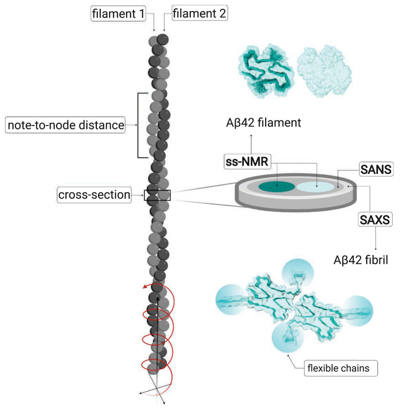
Fig. 1.
Cartoon illustrating a fibril (gray) with its two filaments in light and dark gray. The node-to-node distance can be obtained from AFM or cryo-EM, which can also provide information of the detailed packing. To the Right is shown an excised cross-section plane, the dimensions of which are studied by SAXS and SANS, with the cores of the two filaments in tile and light cyan. Above the cross-section is shown one plane of each filament, seen from the Top, based on the ss-NMR structure (1) PDB ID 5KK3, DOI: 10.2210/pdb5KK3/pdb, with one filament as space-filling model and the other one as space-filling model with the backbone of the two monomers outlined. Below the cross-section is shown one plane of the fibril with the backbone of all four monomers outlined and the flexible N-termini indicated by cyan ovals, as modeled in the current work. (Created with BioRender.com.)

