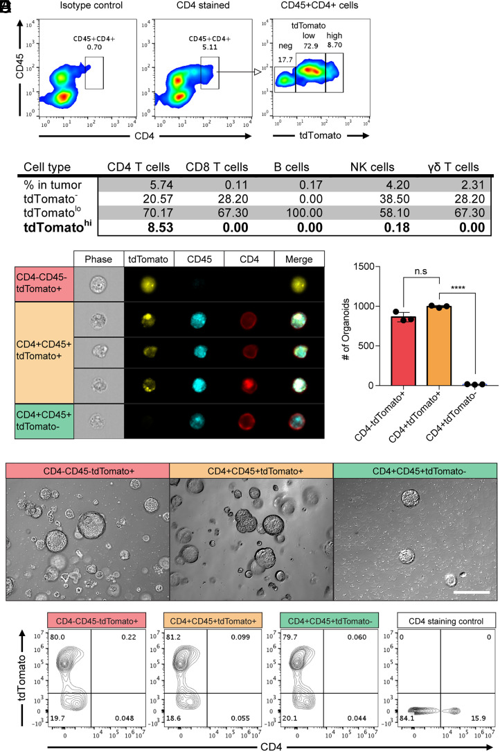
Fig. 1.
Colon cancer cells acquired membrane CD4 from CD4-positive T cells in vivo. Colon cancer organoids (AKPS) carrying mutations in Apc, Kras, Tp53, and Smad4 genes with a tdTomato reporter gene were injected into C57BL/6 mice via splenic vein to develop liver metastases. (A) Surface staining of CD4 and CD45 identified the double-positive cell population. Fluorescence intensity of tdTomato in this population is shown. Isotype control: isotype control of anti-mouse CD4 antibody. The numbers indicate the percentile of each population. (B) A summary of the average percentiles of tdTomato-positive, tumor-infiltrated lymphocytes analyzed using flow cytometry. CD45, CD4, CD8, CD19, NK1.1, and γδ-T cell receptor were stained to identify each cell type. tdTomatolo and tdTomatohigh populations are gated as shown in A. (C) Amnis imaging flow cytometry analysis of the expression and staining patterns of tdTomato, CD4, and CD45 in cells from tumor tissues. (D and E) Generation of colon cancer organoids from the sorted cells based on the expression of tdTomato, CD4, and CD45 (CD4+tdTomato+ cells from the CD45+CD4+tdTomatohigh gating) as described in SI Appendix, Fig. S2. The 2,000 sorted cells were plated in 50 μL Matrigel. The numbers of organoid formation on day 7 after plating the sorted cells (D) and representative pictures (E) are shown. n.s, not significant, ****P < 0.0001; two-tailed t test. Error bars indicate mean ± SD.Scale bar: 50 μm. (F) Expanded organoid cells on day 7 in D and E were harvested and stained with anti-mouse CD4 monoclonal antibody. The intensity of tdTomato and CD4 was analyzed. CD4-positive control cells are spleen cells from the C57BL/6 actin-GFP reporter mice. Results are representative of two to five independent experiments.

