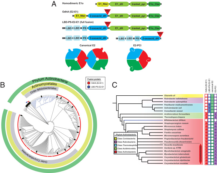
Fig. 5.
Distribution of OdhA orthologs and PCI-bearing E2 enzymes in the Actinobacteria phylum. (A) Schematic representation of the different domain associations. Green blocks indicate Pfam domains associated to homodimeric E1o enzymes, while blue blocks refer to domains composing E2 enzymes. The respective Pfam accession numbers are indicated in the Material and Methods section. LBD: lipoyl binding domain; PS: PSBD. (B) Distribution of OdhA orthologs (red dots) or full E2o-E1o fusion enzymes (LBD-PS-E2-E1; blue dots) in the phylum Actinobacteria. Branches highlighted in black correspond to genomes selected to be analyzed for the PCI presence in E2. (C) Representative sequences from the actinobacterial tree, showing the correlation between the presence of OdhA-like enzymes or full E2o-E1o fusion enzymes, and PCI-bearing enzymes versus canonical E2 enzymes.

