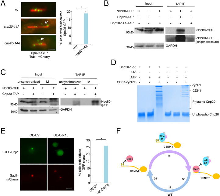
Fig. 7.
The interaction of Ndc80-CENP-T is abolished in the phospho-null CIM mutant and CDK1 phosphorylates the CIM domain. (A) Spc25-GFP localization in the cnp20-14A mutant is aberrant (white arrow) during mitosis. Tub1-mCherry was used as a microtubule marker. Right: Quantification of the percentage of indicated cells with mislocalized Spc25-GFP. (Scale bar, 2 μm.) (B) Lysates from indicated cells synchronized at metaphase using nda3-KM311 were immunoprecipitated with an antibody specific for TAP. Precipitated proteins were analyzed by Western blotting using a GFP antibody. GAPDH was used as a loading control. (C) Cell lysates from cells expressing Ndc80-GFP and CENP-TCnp20-TAP either synchronized in mitosis or unsynchronized were immunoprecipitated with an antibody specific for TAP. Precipitated proteins were analyzed by Western blotting using indicated GFP antibody. Cells expressing Ndc80-GFP were used only as a control. GAPDH was used as a loading control. (D) In vitro kinase assays were performed using human recombinant CDK1-cyclin B and a synthesized peptide derived from CENP-TCnp20 1 to 55. The CENP-TCnp20 1 to 55-14A peptide (14A) was used as a control. Samples were analyzed by Phos-tag PAGE. (E) Constitutive overexpression of Cyclin B/Cdc13 results in dissociation of Ccp1 from centromeres. Cells carrying pREP1-empty vector (pREP1-EV) or pREP1-cdc13 were incubated on the minimal pombe minimal glutamate (PMG) medium without thiamine at 30 °C for 22 h. Sad1-mCherry was used as an SPB marker. (Scale bar, 2 μm.) Right: Quantification of the percentage of indicated cells with diffuse GFP signal in the nucleus. Experiments were performed in triplicate. At least 40 cells were scored in one single experiment. Error bars represent mean and SD. *P < 0.05. (F) Model: the CIM domain of CENP-T is phosphorylated at the onset of mitosis by CDK1, which dissociates Ccp1 from CENP-T, allowing proper positioning of Ndc80C at the adjacent Ndc80 receptor motif. At the end of mitosis, the CIM domain is dephosphorylated by an unknown phosphatase. This leads to the reassociation of Ccp1 with the CIM domain.

