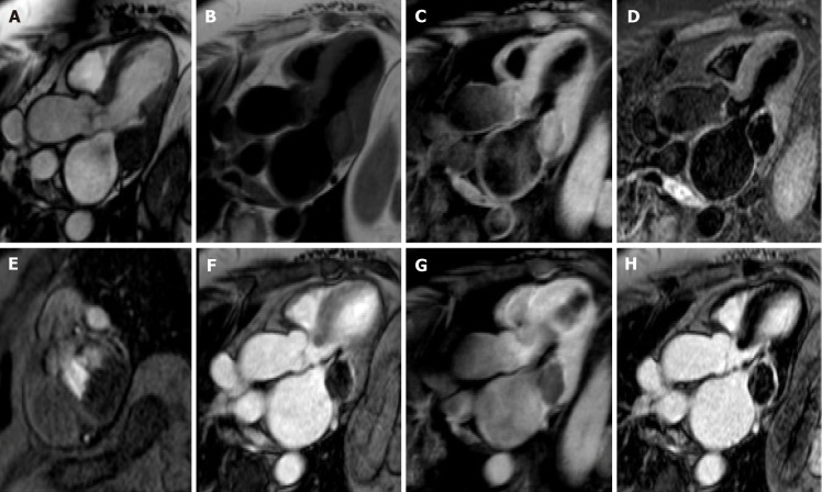
Figure 6.
Eighty-year-old female with hyperechoic mass of uncertain significance in the left atrio-ventricular groove discovered at transthoracic echocardiography. Cardiovascular magnetic resonance confirms the presence of the mass with the cine- steady state free precession images (A). The mass shows slight hyperintense signal on T1-weighted images without and with fat suppression (B and C, respectively), due to the presence of proteinaceous material, and hypointense signal on short tau inversion recovery images (D). The mass shows no contrast uptake during perfusion sequences (E) and no contrast enhancement both on early gadolinium enhancement images (F), T1- weighted images (G) repeated after the contrast medium injection, and late gadolinium enhancement images (H) with a peripheral rim of enhancement. These findings are in keeping with caseous calcification of the mitral valve.

