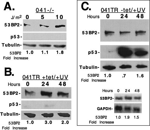
FIG. 4.
p53-independent mechanisms participate in the UV irradiation induction of 53BP2. (A) Western blot of lysates (15 μg of total protein per lane) prepared from 041−/− cells 24 h after UV irradiation with 0, 5, or 10 J/m2. The fold increase of 53BP2 protein compared to no-UV-irradiation lysates are indicated below, normalized for tubulin. (B) Western blot of equivalent amounts of lysates prepared from 041TR cells maintained in 2 μg of tetracycline per ml at the indicated times after UV irradiation at 20 J/m2. The fold increases of 53BP2 protein compared to the levels at time zero are indicated below, normalized for tubulin. (C) The upper panels show Western blots of equivalent amounts of lysates prepared from 041TR cells at the indicated times after tetracycline withdrawal. At time zero, cells were UV irradiated with 20 J/m2. The fold increases of 53BP2 protein compared to levels at time zero are indicated below, normalized for tubulin. 53BP2 protein was detected with monoclonal antibody DX547; p53 was detected with monoclonal antibody 1801. The lower panels show Northern blots of total RNA (15 μg/lane) prepared from the 041TR cells above at the indicated time points, probed for 53BP2, stripped, and reprobed for GAPDH. The fold increases of 53BP2 mRNA compared to the levels at time zero are indicated below, normalized for GAPDH.

