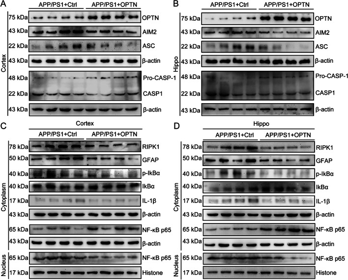
Fig. 10.
Overexpression of OPTN in the brains of APP/PS1 transgenic mice alleviates activation of the AIM2 inflammasome and RIPK1 pathways. A–D Three-month-old APP/PS1 transgenic mice were injected with OPTN or control adeno-associated virus in the hippocampus and cortex for 1 month. Brain tissue was collected after anesthesia euthanasia. A, B Western blotting was used to detect the protein expression of OPTN, AIM2, ASC and caspase-1 in the cerebral cortex and hippocampus of mice. β-actin served as the internal control. C, D Western blot analysis was used to determine levels of RIPK1, GFAP, p-IKBα, IKBα, IL-1β, and NF-κB in the cytoplasm and nucleus of mice. Histone and β-actin were used as internal controls for the nucleus and cytoplasm, respectively. The data present means ± S.M. of independent experiment. OPTN-AAV injected APP/PS1 Tg mice compared with control-AAV injected APP/PS1 Tg mice, *P < 0.05, **P < 0.01, ***P < 0.001

