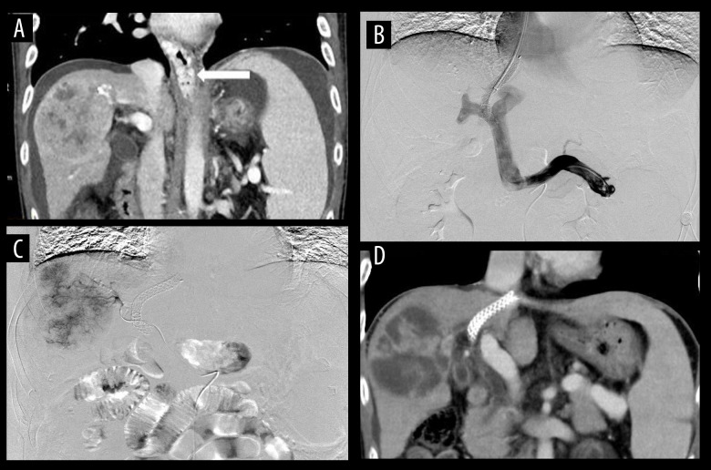
Figure 2.
(A) The examination of esophageal and gastric varices (white arrow), peritoneal effusion, and hepatocellular carcinoma by computed tomography (CT) in patients. (B) The effective shunt function of the stent through angiography under transjugular intrahepatic portosystemic shunt (TIPS), with implantation of 8×70-mm Viatorr stent. (C) In patients undergoing transarterial chemoembolization (TACE), the staining of hepatocellular carcinoma was obvious and embolization with drug-loaded microspheres was performed. (D) At the 1-month follow-up after TACE, most of the hepatocellular carcinoma was necrotic, the stent was unobstructed, and the peritoneal effusion disappeared.

