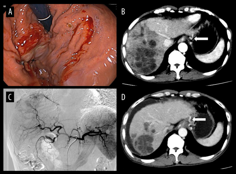
Figure 3.
(A) Gastroscopy revealed varices in the fundus of the stomach, which showed bleeding and rupture of blood vessels. Under the gastroscope, application of titanium clips to hemostatic varicose veins and tissue glue injection were used to stop the bleeding. (B) The hepatocellular carcinoma and gastric fundus varices (white arrow) by computed tomography examination. (C) In patients undergoing transarterial chemoembolization (TACE), the staining of hepatocellular carcinoma was obvious and embolization with drug-loaded microspheres was performed. (D) At the 1-month follow-up after TACE, most of the hepatocellular carcinoma was necrotic, but there was peritoneal effusion, and the gastric fundus varices were still not relieved (white arrow).

