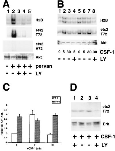
FIG. 3.
An Akt immunoprecipitate catalyzes phosphorylation of ets-2 at position threonine 72, and LY294002 inhibits ets-2 phosphorylation in me-v macrophages. (A) Akt immune kinase assays performed with RAW264 cells using histone H2B, ets-2 T72, and ets-2 A72 as substrates (panels as indicated). Akt was isolated from 107 cells grown in normal medium (lane 1), treated for 5 min with 300 μM pervanadate (pervan) (lane 2), or treated for 5 min with both 300 μM pervanadate and 50 or 100 μM PI 3-kinase inhibitor LY294002 (LY) (lanes 3 and 4, respectively). The sample in lane 5 is a control in which Akt antibody was not included. The bottom panel is a Western blot performed with the Akt antibody to demonstrate that Akt was present in all samples. (B) Akt immune kinase assays performed on 107 wild-type (lanes 1 to 4) or me-v primary macrophages (lanes 5 to 8) with histone H2B or ets-2 substrates (as labeled). Cells were grown without CSF-1 for 24 h and then stimulated with 50 ng of CSF-1 per ml for the times indicated. LY294002 (100 μM) was included 30 min prior to addition of CSF-1 (lanes 4 and 8). The bottom panel is a Western blot of the immunoprecipitated material probed with anti-Akt antibody. (C) The average of four independent Akt immune kinase assays performed on wild-type (WT [open bars]) or me-v (shaded bars) macrophages with the ets-2 substrate (including the experiment shown in 3B). The results are presented relative to wild-type samples grown in the absence of CSF-1. The error bars indicate the standard deviation. (D) Western analysis of wild-type (lanes 1 and 2) or me-v (lanes 3 and 4) macrophages by using the anti-ets-2 pT72-specific antibody (top panel). Cells were grown in the presence of 50 ng of CSF-1 per ml and treated with 100 μM LY294002 for 16 h (lanes 1 and 4). The blot shown was reprobed with an anti-Erk antibody as a sample loading control (bottom panel).

