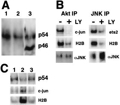
FIG. 4.
p54 JNK coimmunoprecipitates with Akt and is an ets-2 kinase in me-v macrophages. (A) Akt immunoprecipitates (lanes 1 and 2) were analyzed by Western blotting with the anti-JNK antibody. Cells were grown in the absence of CSF-1 for 24 h (lanes 1) or continuously in the presence of 50 ng of CSF-1 per ml (lanes 2). Whole-cell extracts were also prepared and analyzed on a lane on the same gel (lane 3). The positions of the p54 and p46 JNK isoforms are indicated. (B) Akt (left panels) or JNK (right panels) immune kinase assays performed on me-v macrophages using N-terminal c-jun, ets-2 “pointed,” and histone H2B substrates, as indicated. Cells were grown continually in the presence of 50 ng of CSF-1 per ml and treated with 100 μM LY294002 (LY) for 16 h as indicated. The bottom panel is a Western blot with an anti-JNK antibody to demonstrate that equivalent amounts of JNK were present in untreated and LY294002-treated samples. IP, immunoprecipitate. (C) Wild-type BMMs were deprived of CSF-1 for 24 h (lane 1) and then restimulated with CSF-1 for 5 or 30 min (lanes 2 and 3, respectively). Cell extracts were prepared and incubated with anti-Akt antibody. Half of the Akt immunoprecipitate was analyzed by Western blotting with a JNK antibody (top panel, p54). The other half was assayed for kinase activity by using both the c-jun (middle panel) and histone H2B substrates (lower panel).

