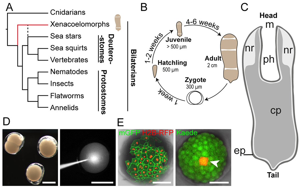
Figure 1: Hofstenia miamia is a highly regenerative species with accessible and manipulable embryos.

(A) Simplified phylogenetic tree of metazoans showing placement of Hofstenia within xenacoelomorphs (red line). Dashed black line indicates the alternative position of xenacoelomorphs, as sister group to ambulacrarians (e.g., sea stars); (Philippe et al., 2019; Kapli and Telford, 2020; Kapli et al., 2021). (B) Life cycle of Hofstenia miamia in lab conditions. (C) An adult worm schematic drawing showing major structures: m, mouth; nr, neural condensation (Hulett, Potter and Srivastava, 2020); ph, pharynx; cp, central parenchyma; ep, epidermis. (D) Hofstenia embryos are amenable to microinjections. Left, live embryos at 2-cell and zygote stages; right, injection of fluorescent dextran into a zygote. Scale bars, 200 μm. (E) Translation of foreign mRNAs. Left, detection of nuclear-localized H2B-RFP and membrane-bound GFP (mGFP), 1.5 days after single blastomere injection of a 2-cell stage embryo; right, single cell photoconversion of Kaede (white arrowhead), 2 days after injection of Kaede mRNA into a zygote. Scale bars, 100 μm.
