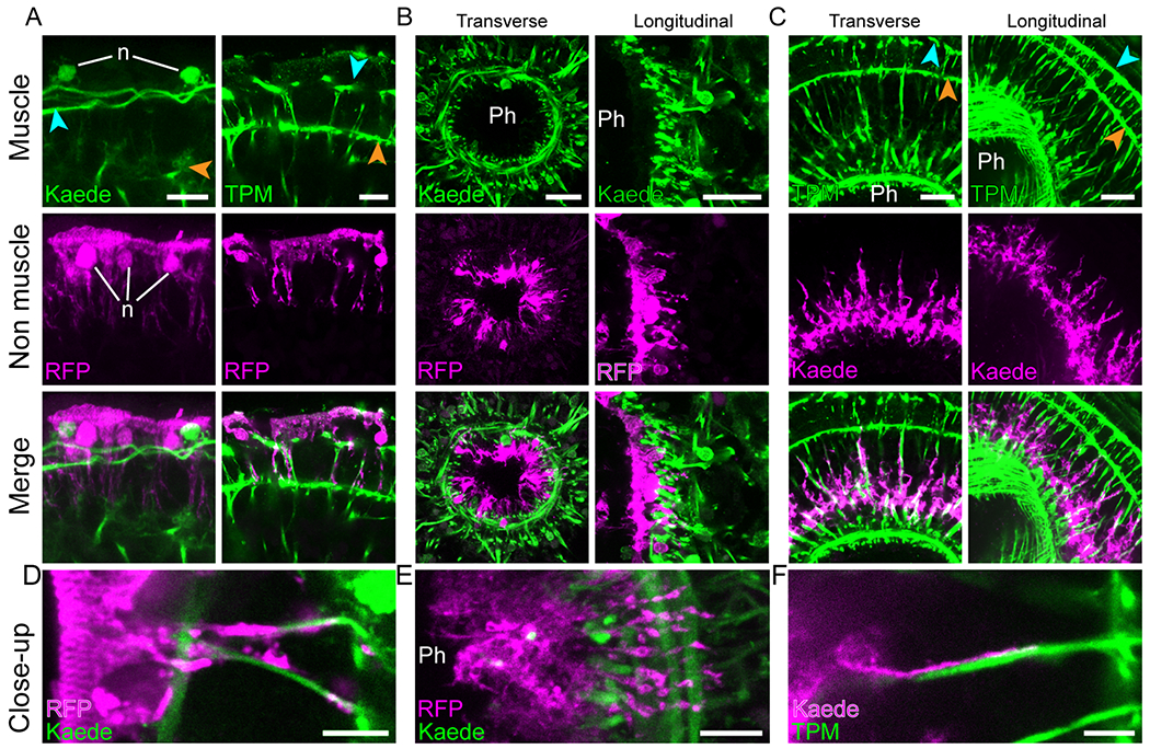
Figure 5: Muscle fibers closely contact epidermal, pharyngeal, and digestive cells.

Dual labeling of muscles with epidermal (A, D), pharyngeal (B, E) and digestive (C, F) tissues. (A) Interaction of epidermal cells with peripheral muscle fibers (left) and ramifications of parenchymal muscle fibers (right). (B) Pharyngeal cells projecting ramifications through pharyngeal muscle net. Cross (left) and longitudinal (right) sections of the pharynx. (C) Digestive tissue projecting ramifications along parenchymal muscle fibers as seen in cross (left) and longitudinal (right) sections. (D, E and F) High magnification images showing the close interaction between muscle fibers and epidermal, pharyngeal and digestive cell ramifications, respectively. Blue and orange arrowheads point to peripheral and body wall muscles, respectively. Images are in pseudo-colors with green indicating muscle detected either as Kaede fluorescence or anti-Tropomyosin immunolabeling (TPM) and magenta indicating epiA+ or psap+ cells detected either as Kaede fluorescence or TagRFP-T immunolabeling (RFP); detection method indicated at bottom left corner. Except for TPM, fluorescent labeling originates from transgene expression. Images are representative of at least ten samples. Ph, pharynx lumen; n, nuclei. Scale bars, A, 10 μm; B and C, 20 μm; D, E, F, 5 μm. See also figure S5 and video S7.
