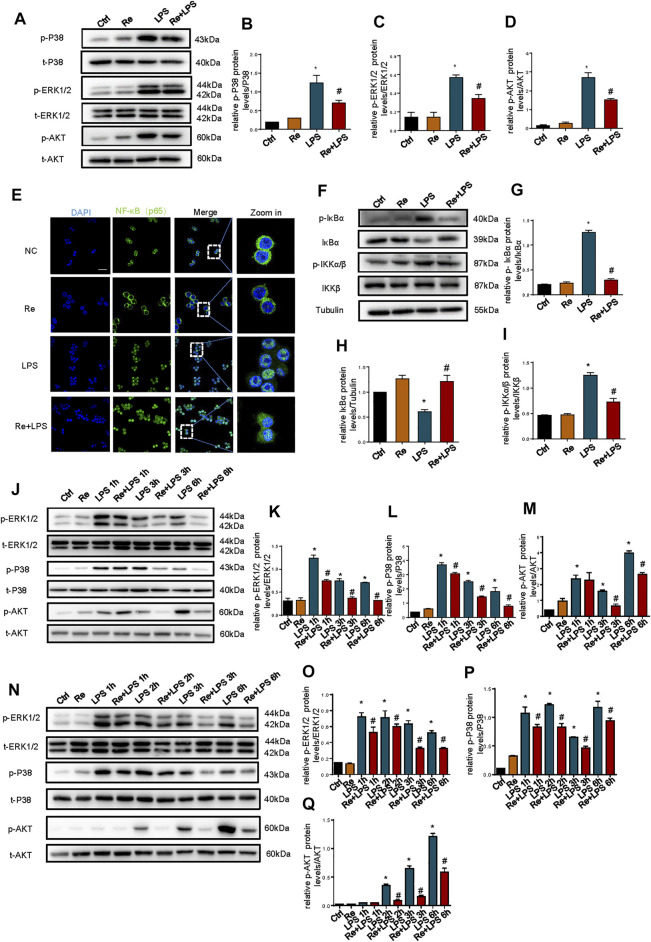
FIGURE 3.
Suppressive effects of remimazolam on LPS induced activation of the MAPK and PI3K signaling pathway in macrophage. Macrophages were treated with LPS (1 μg/ml) with or without pretreatment of remimazolam (Re). (A) Western blot analysis of p-P38, P38, p-ERK1/2, ERK1/2, p-AKT, and AKT protein expression were measured 15 min after LPS treatment in RAW264.7 cells. (E) Immunofluorescence staining of NF-κB p65 in RAW264.7 were measured 6 h after LPS treatment. (F) Western blot analysis of IκBα, p-IκBα, IKKα/β, and p-IKKα/β protein expression were measured 15 min after LPS treatment in RAW264.7 cells. (G–I) Protein presence of phosphorylated IκBα, IκBα, and phosphorylated IKKα/β was normalized to IκBα, β-tubulin, IKKα/β respectively. (J) Western blot analysis of p-ERK1/2, ERK1/2, p-P38, P38, p-AKT, and AKT protein expression were measured 1, 3, 6 h respectively after LPS treatment in Raw264.7 cells. N Western blot analysis of p-ERK1/2, ERK1/2, p-P38, P38, p-AKT and AKT protein expression were measured 1, 2, 3, 6 h respectively after LPS treatment in BMDM. (B–D), (K–M), (O–Q) Protein presence of p-ERK1/2, p-P38, and p-AKT was normalized to ERK1/2, P38, and AKT respectively. Each bar represents the mean ± SD. (n = 3), *p < 0.05, compared with negative control group; #p < 0.05, compared with LPS group.

