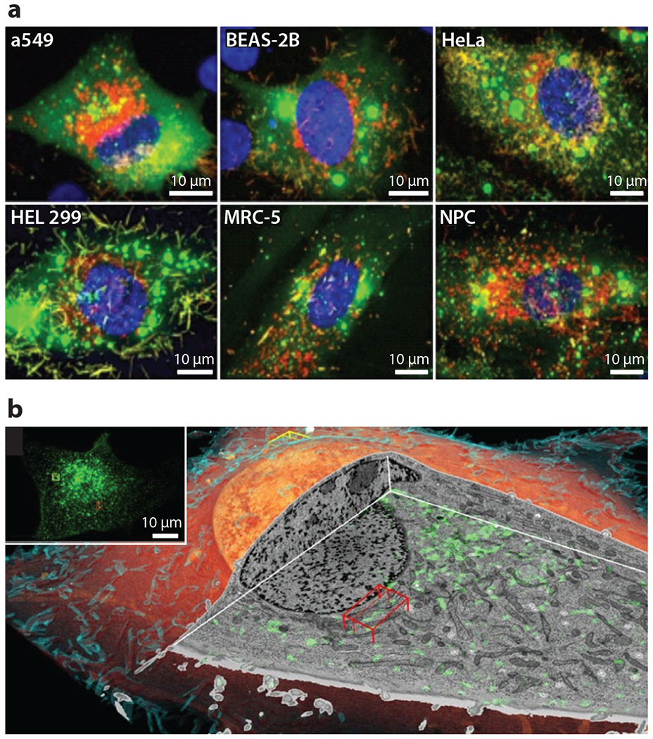
Figure 5.

Fluorescence and correlative microscopy. (a) Fluorescence light microscopy using spinning-disk confocal and laser scanning confocal to determine the permissivity of cell lines to RSV. The images are stained for RSV fusion protein (red), nucleocapsid (green), and nucleus (blue). Adapted with permission from Reference 35. (b) Volume rendering of cryo-FIB milling coupled to SEM of a SUM159 cell. The inset is a correlative cryo–three-dimensional SIM image where transferrin is colored green to show the endolysosomal compartments. Abbreviations: FIB, focused ion beam; RSV, respiratory syncytial virus; SEM, scanning electron microscopy; SIM, structured illumination microscopy. Adapted with permission from Reference 38.
