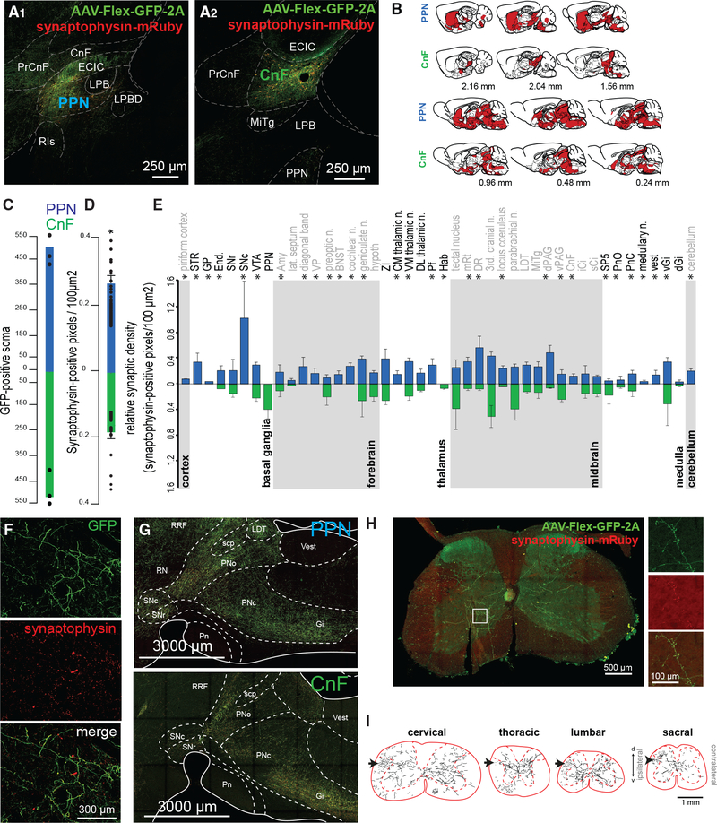
Figure 2. Axonal distribution of PPN and CnF glutamatergic neurons.
(A and B) Following injection of AAV-DIO-GFP-2A-synaptophysin-mRuby limited to the PPN (A1) or the CnF (A2) borders based primarily on ChAT staining (A1′ and A2′), we observed widespread distribution of GFP-labeled axons across the brain (sagittal plane) (B).
(C and D) Quantification of the total count of GFP-positive soma (PPN: 504.66 ± 58.42, CnF: 518.33 ± 54.67; one-way ANOVA, F(1,5) = 0.03, p = 0.87) and overall synaptic density (PPN: 0.27 ± 0.024 pixels/100 μm2, CnF: 0.18 ± 0.02 pixels/100 μm2; one-way ANOVA, F(1,86) = 7.64, p = 0.007).
(E) Segregated synaptophysin labeling across the brain revealed distinct patterns of innervation by PPN and CnF glutamatergic neurons, particularly in the basal ganglia, forebrain, thalamus, midbrain, medulla, and cerebellum (Wilcoxon test).
(F) Fluorescent micrographs illustrating a representative example of GFP and synaptophysin labeling in the striatum following PPN transduction.
(G) Distribution of axons in the brainstem following PPN and CnF injections (sagittal plane).
(H) Synaptic distribution in a cervical segment of the spinal cord.
(I) Axonal reconstructions in typical examples of cervical, thoracic, lumbar, and sacral spinal cord segments following unilateral PPN injection. Black arrows represent the rubrospinal tract.
*p < 0.05. All experiments have been replicated in at least 3 mice. A single datum is represented by a small dot. All data are represented as mean ± SEM. Scale bars: (A) 250 μm, (A′) 50 μm, (F) 300 μm, (G) 3,000 μm, (H) 500 μm, (H, inset) 100 μm, (I) 1 mm.

