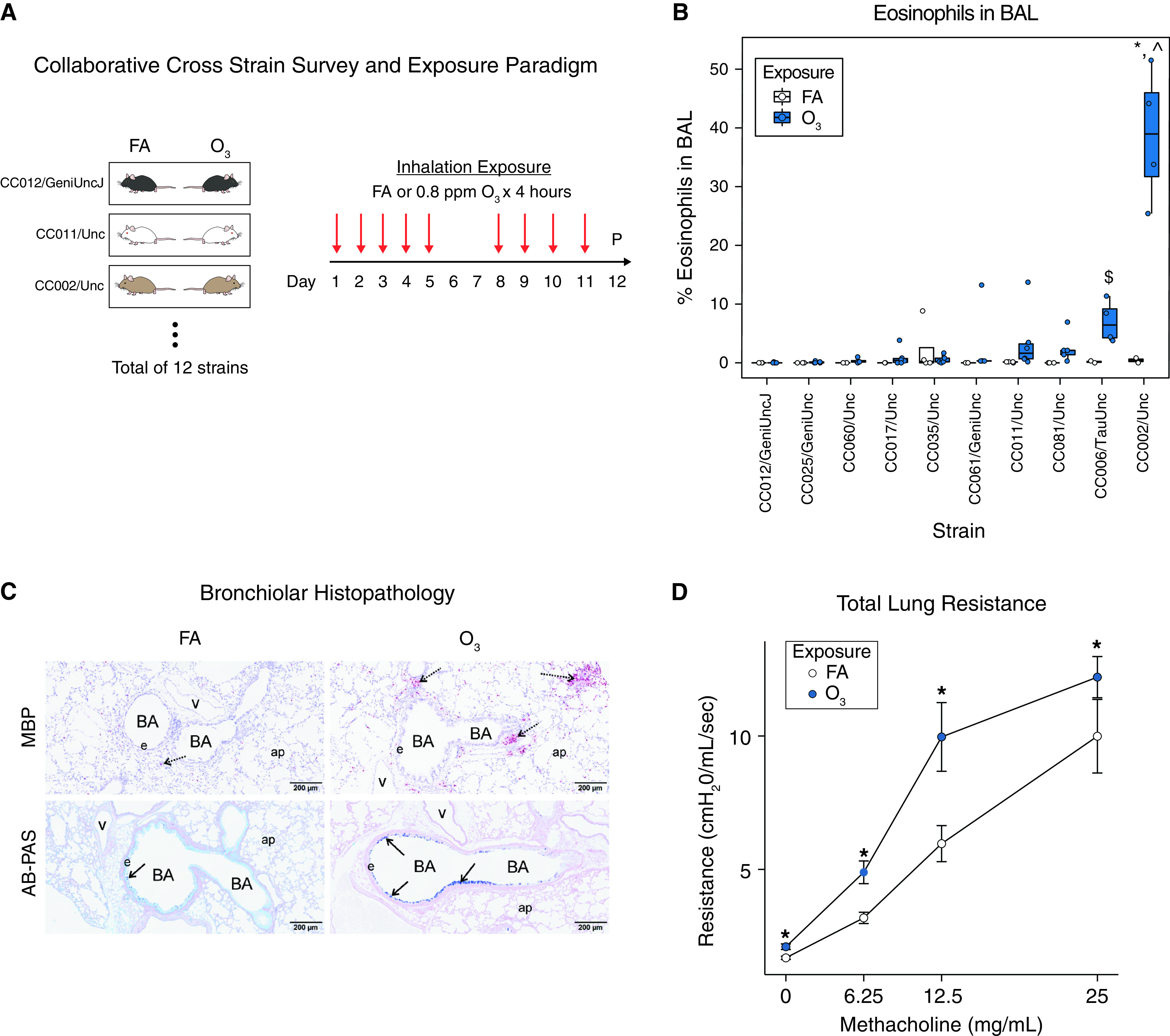
Figure 1.

Repeated O3 exposure causes eosinophilic inflammation, airway mucous cell metaplasia, and airway hyperresponsiveness in CC002 mice. (A) Experimental design involving repeated exposure to O3 or filtered air (FA) across 12 Collaborative Cross (CC) strains. Mice were phenotyped 21 hours after last exposure. (B) Variation in O3-induced airway eosinophils in BAL fluid in 10 out of 12 CC strains tested. See Supplement for data on other cell types and CC strains. *P < 0.05 and $P < 0.01 for O3 versus FA; ⁁P < 0.05 versus other strains. FA, N = 2–4 per strain; O3, N = 4–6 per strain. (C) Representative histological sections of FA controls and O3 exposed CC002 mice showing (top) peribronchial eosinophilic inflammation, detected by immunohistochemical staining for major basic protein (MBP, red stain), and (bottom) mucous cell metaplasia in bronchiolar epithelium as detected by Alcian blue–periodic acid–Schiff (AB-PAS) staining. Scale bars, 200 μm. BA = bronchial airway, e = epithelium, ap = alveolar parenchyma, v = blood vessel. Arrows denote regions of airways featuring eosinophils or mucous cells. (D) Total lung resistance at baseline and after escalating doses of methacholine in O3 exposed (vs. FA control) CC002 mice in a separate experiment. N = 7 per group. *P < 0.05 versus FA control.
